Ceramics play an important role in our lives. Researchers have now found an insight into building stronger and tougher ones by studying shells of bivalve molluscs. This was done by Ling Li, an assistant professor in mechanical engineering at Virginia Tech.
The study was formed by examining the capacity of basic mineral building blocks in the shell to expect fractures, instead of focusing only on the shape and chemistry of the structure.
The team conducted an in-depth analysis on microscopic structures of the shells of pen shell molluscs, bivalves native to the Caribbean. These shells comprise two layers, an inner nacre layer and a brown-coloured outer layer. The inner layer is also known as mother-of-pearl which is often iridescent because of its regular nanoscopic layering structure, similar to the colouration mechanism for many bottlefly wings, whereas the outer layer is composed of prism-shaped calcite crystals arranged in a mosaic pattern.
Between adjacent mineral crystals, very thin (approximately 0.5 micrometers, less than one-hundredth the size of a human hair) organic interfaces are present that glue the crystals together. The calcite crystals measure approximately half a millimeter in length and 50 micrometers in diameter, resembling elongated prisms.
The calcite crystals, unlike other synthetic crystals, contain many nanoscopic defects, primarily composed of organic substances.
Li added that we can consider the biological ceramic, in this case, the pen shells’ calcite crystals, as a composite structure, where many nanosized inclusions are distributed within its crystalline structure. The calcite shell itself is a single crystal shell, which makes this remarkable. A presence of structural defects means there is a site of potential failure.
This is the reason the normal approach to minimize the structural discontinuities or stress concentrations in engineering structures fails.
Li’s team to the contrary showed the size, spacing, geometry, orientation, and distribution of these nanoscale defects within the biomineral is highly controlled, improving not only the structural strength but also the damage tolerance through controlled cracking and fracture.
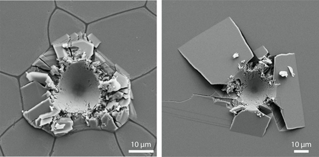
When an external force is applied on these shells, the crystal minimized plastic yielding by impeding the dislocation motion, a common mode for plastic deformation in pure calcite, aided by those internal nanoscopic defects.
This design besides adding strength allows the structure to use its crack patterns to minimize the damage to the inner shell. The mosaic-like interlocking pattern of the calcite crystals in the prism layer further contains large-scale damage when the external force is spread across the individual crystals. The structure can crack to dissipate the external loading energy without failing.
Li concluded by adding that these nanoscopic defects aren’t some random structure but play an important role in the mechanical properties of this natural ceramic. This research has helped them to know the organism really turns the originally weak and brittle calcite to a durable biological armour. They are now focusing on fabrication processing, such as 3D printing, to implement these strategies to develop ceramic composites with enhanced mechanical properties for structural applications.
Journal Reference:
Zhifei Deng, Hongshun Chen, Ting Yang, Zian Jia, James C. Weaver, Pavel D. Shevchenko, Francesco De Carlo, Reza Mirzaeifar, Ling Li. Strategies for simultaneous strengthening and toughening via nanoscopic intracrystalline defects in a biogenic ceramic. Nature Communications, 2020; 11 (1) DOI: 10.1038/s41467-020-19416-2
Press Release: Virginia Tech