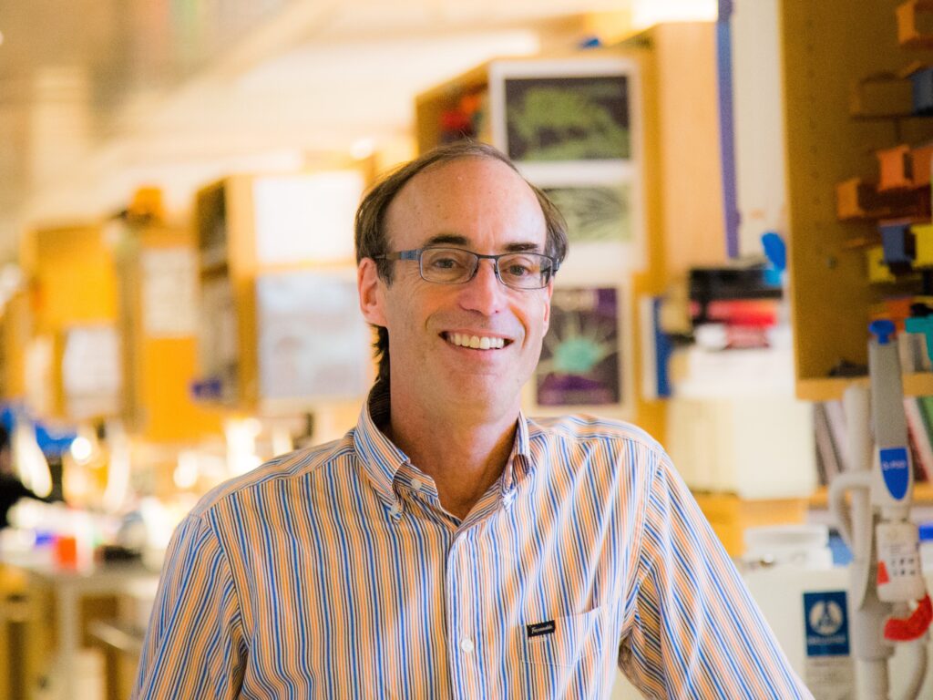
Ronald Vale is a well-known American cell biologist and a biochemist, known for his work on motor proteins, in particular kinesin and dynein.
He is currently a professor at the Department of Cellular Molecular Pharmacology, University of California, San Francisco. He has also been an investigator at the Howard Hughes Medical Institute since 1995. In 2020, Vale became executive director of the Janelia Research Campus and a vice president of HHMI.
He is involved in several science projects through which he tries to improve science education and research.
Index
Education
Ronald Vale was born in Hollywood, California on January 11, 1959. He completed his schooling from Hollywood High School. He earned his bachelor’s degree in chemistry and biology from the College of Creative Studies, University of California, Santa Barbara in 1980.
He joined an MD/Ph.D. program at Stanford University, where he earned his Ph.D. in neuroscience under Eric Shooter while working with the Nerve Growth Factor.
He had also worked in the laboratories of C. Fox at UCLA and Robert Lefkowitz at Duke University, during the period of his study. He did not finish his MD and joined the University of California, San Francisco as an assistant professor in 1986, and became a full professor in 1994.
Scientific Research

The Nerve Growth Factor is a neuropeptide (peptides are small chains of amino acids) which is also a neurotrophic factor. It is able to signal and hence drive or regulate certain aspects of neurons (the building block of the nervous system). They are responsible for the regulation of growth, maintenance, survival, etc. of the neurons.

As Ronald Vale worked on the Nerve Growth Factor he began to be interested in the mechanisms behind the transport of molecules and receptors in the nerve axon. Michael Sheetz and James Spudich had then shown myosin moving along the actin filaments. Vale hypothesized that this movement was the mechanism behind the movement of organelles in the axon. Myosin is a motor protein. Motor proteins are proteins that can ‘motor’ (move) through the cytoplasm. Some motor proteins move along the cytoskeletal elements.
Wanting to work on this hypothesis, Vale and Sheetz collaborated with Bruce Schnapp and Tom Reese, at Reese’s laboratory at the Marine Biological Laboratory, where they worked on the giant squid axon. They discovered that organelle transport happened bidirectionally on microtubules instead of actin filaments. They also isolated from the cytosol, a motor protein that they named kinesin. Kinesin was seen to move in only one direction of the microtubule, and later on, dynein (discovered by Ian Gibbons in 1965; function given by Richard Vallee) was seen to move in the opposite direction.
Vale discovered that purified organelles seldom moved on microtubules, unless in the presence of axonal cytosol. He also discovered that microtubules moved on glass surfaces in the presence of cytosol, as well as cytosol covered beads moved on the microtubules. This allowed him to elucidate assays to study microtubule-based motility. An assay is any procedure used to qualitatively or quantitatively measure the amount, function, etc. of a protein or other molecules.
Vale along with Jonathan Howard and A. James Hudspeth developed a single molecular assay for kinesin in 1989. Later on, in 1996, Vale with Toshio Yanagida created a single-molecule fluorescence assay for kinesin. He also discovered the first protein to break apart microtubules in the year 1991, naming it katanin. In 1996, Vale and his team solved the crystal structure of the kinesin motor domain, and later on went to suggest a model for the mechanism of kinesin movement on the microtubule.
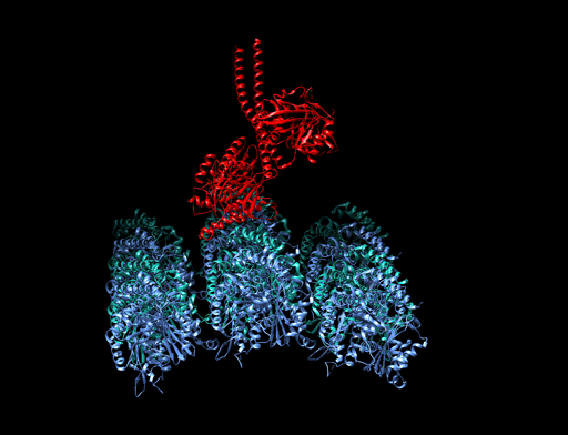
Since 2003, Vale has worked on the dynein motor protein. Apart from determining the structure of the dynein microtubule-binding domain and the dynein motor domain, Vale’s laboratory has also elucidated the movement of dynein on microtubules.
Helping Science Education and Research
Ronald Vale founded iBiology (ibiology.org) in 2006, which is a non-profit organization that aims to improve biology education by releasing free online videos and courses on various biological research, all made by leading biologists.
He also set up the Young Investigators’ Meeting in India in 2009, which provides an opportunity for postdocs and junior fellows in India to network, and receive mentoring.
He co-founded the ASAPbio (Accelerating Science and Publication in Biology) (asapbio.org) in 2015. The organization’s objective is to promote an open and transparent peer-review process as well as the use of preprints, among other tasks and aims.
He also founded Indiabioscience (Indiabioscience.org) which is a non-profit science outreach initiative created to fulfill the niche gap within the Life Science sector in India. It promotes the Life Sciences in India, by enabling networking, being an information hub, promoting skills, and communicating science.
He is also an organizer of the Bangalore Microscopy Course. This course provides didactic and hands-on training in state-of-the-art optical microscopy techniques. A range of topics from basic microscopy to super-resolution imaging is covered. Research seminars detailing applications of light microscopy to address specific biological questions is also a part of the program.
Honors
He’s been awarded
- Wiley Prize in Biomedical Sciences 2012
- Albert Lasker Award for Basic Medical Research in 2012
- Shaw Prize in Life Science and Medicine in 2017
- Canada Gairdner International Award for Biomedical Research in 2019, among many other awards.
Vale is a fellow of the American Academy of Arts and Sciences as well as a foreign fellow of the Indian National Science Academy. He’s a member of the National Academy of Sciences and was the president of the American Society for Cell Biology in 2012.